Friedreich Ataxia Patients Have Atypical Brain Makeup, Study Says
Written by |
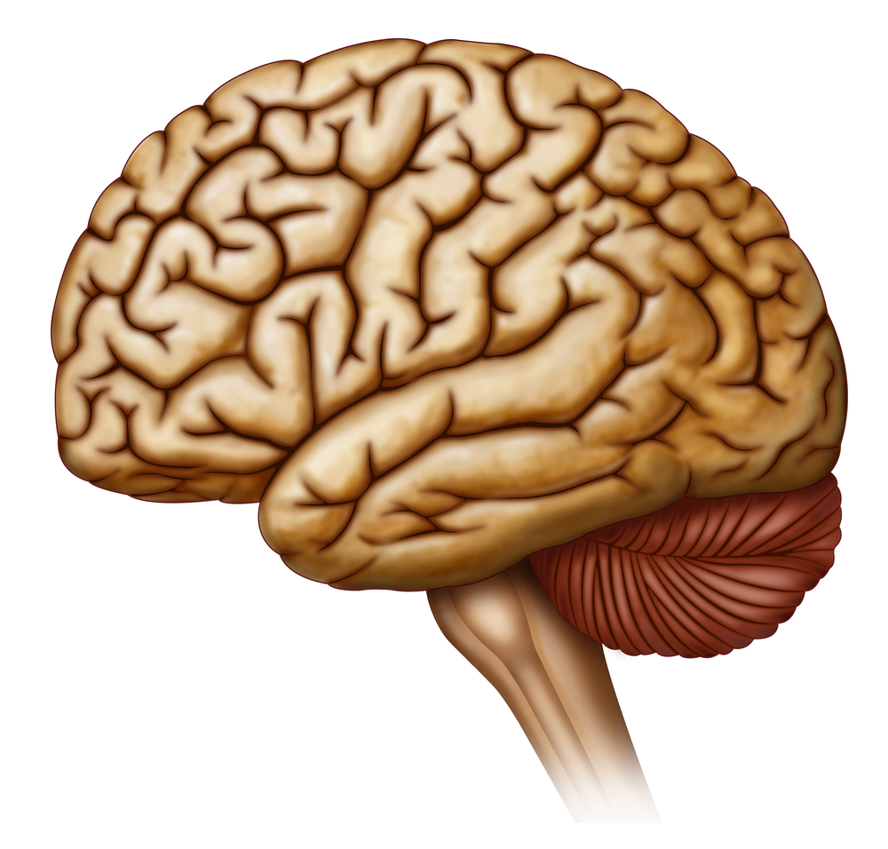


Several Australian institutions collaborated to evaluate the structural integrity of a major component of the central nervous system, cortical grey matter, in patients with Friedreich ataxia (FRDA). The scientists found structural abnormalities in several brain regions, primarily in the cortical motor system, which represents a step forward in understanding the disease.
The study, titled “Cerebral and cerebellar grey matter atrophy in Friedreich ataxia: the IMAGE-FRDA study”, was published in Journal of Neurology.
In FRDA, the nerve cells responsible for coordination and muscle contraction become damaged over time, which reduces muscle length and creates difficulties in movement. It is believed the disease is caused by genetic mutation, but evidence of cerebral involvement also is growing.
In this study, the researchers evaluated the structural integrity of part of the central nervous system involved in movement, cortical grey matter, in FRDA patients.
A total of 31 FRDA patients and 37 healthy controls were included in the study. The participants were subjected to magnetic resonance imaging to measure thickness and cortical volume in cerebral motor regions-of-interest. The data were then analyzed and the scientists investigated correlations between cortical thickness/volume measures with disease severity, genetic abnormalities, and finger-tapping behavioral measures.
The results suggested that when compared to control participants, FRDA patients showed declined cortical thickness in the motor areas and other regions of the brain. This means that atrophy affects particularly the premotor areas of the cortical motor system, then expands to non-motor areas of the brain.
Other data revealed that volume loss correlates well with measures of clinical severity, genetic abnormality, and motor dysfunction. This suggests that cortical thickness and volume results are divergent, indicating that each of them is sensitive to various aspects of neuropathology in FRDA.
“This study supports the existence of structural abnormalities in the cerebral and cerebellar cortices in FRDA. Tissue atrophy is preferentially observed in pre-motor areas of the cortical motor system, alongside a number of ’hub’ regions of non-motor networks,” the authors concluded.
“Overall, this work adds to a growing literature illustrating pathology in regions distal from the cerebellum and spinal cord, thereby representing a move toward a more complete model of neuropathology in FRDA,” they added.