
Restoration of Frataxin in Neurons from FA Patients May Halt Disease Progression
Written by |


Recent research suggests that correcting the frataxin deficiency in neurons derived from Friedrich’s ataxia (FA) patients may not only stop disease progression, it may also lead to clinical improvement by rescuing dysfunctional surviving neurons.
The study, “Friedreich ataxia induced pluripotent stem cell-derived neurons show a cellular phenotype that is corrected by a benzamide HDAC inhibitor,” was conducted by researchers from Italy and Belgium and published in the journal Human Molecular Genetics.
Friedrich’s ataxia is characterized by reduced levels of the protein frataxin, which contributes to the synthesis of iron-sulfur proteins in mitochondria, the cell’s powerhouse responsible for energy. Low levels of frataxin limit the production of iron-sulfur proteins, which is associated with reduced energy production, oxidative damage, altered iron metabolism, and general mitochondrial dysfunction.
The study’s goal, therefore, was to investigate what would happen when frataxin levels were restored in cells with frataxin deficiency. The researchers used the benzamide HDAC inhibitor 109, which inhibits a group of enzymes that silence gene expression, thereby inducing expression of the frataxin gene.
This compound has been shown to restore frataxin levels in cellular and animal models of the disease, as well as in blood cells from Friedrich’s ataxia patients.
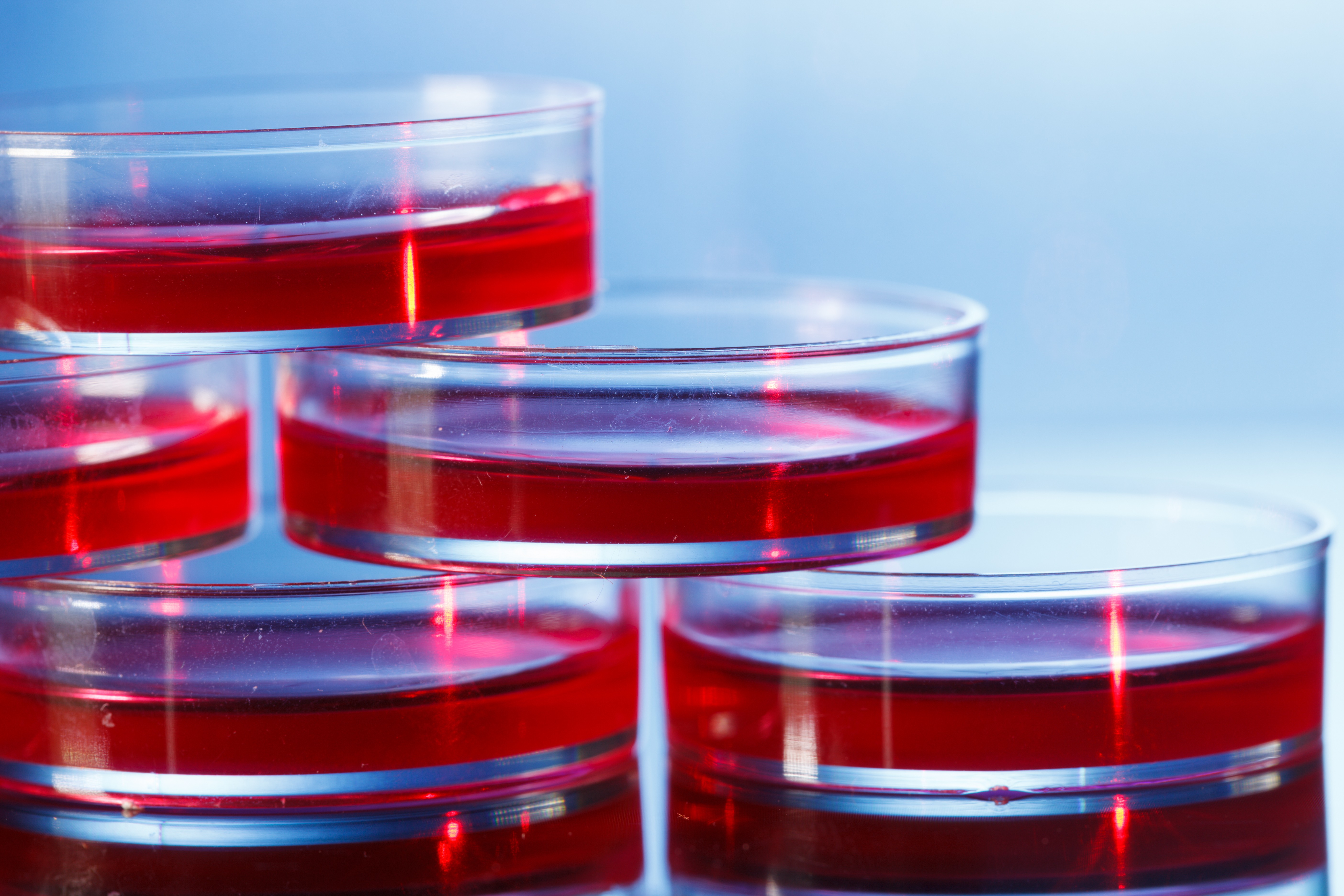
Researchers took skin samples from two Friedrich’s ataxia patients and two healthy controls, and used laboratory techniques that allowed them to reprogram these skin cells to become neurons. In this way, researchers can have cultures of neurons similar to those present in the FA brain and study the molecular changes that underlie this pathology.
As expected, Friedrich’s ataxia neurons showed signs of altered iron metabolism, oxidative stress and deficient iron-sulfur analysis, as indicated by lower levels of iron-sulfur and lipoic acid-containing proteins, higher labile iron pool (LIP), higher expression of mitochondrial superoxide dismutase (SOD2), increased reactive oxygen species (ROS), and lower reduced glutathione (GSH) levels, and enhanced sensitivity to oxidants.
However, treatment with compound 109 significantly increased the levels of frataxin, iron-sulfur and lipoic acid-containing proteins, but decreased SOD2 levels and normalized LIP and ROS levels. It protected Friedrich’s ataxia neurons almost completely against oxidative stress-induced death.
“Taken as a whole, our findings define a robust, reproducible and quantifiable phenotypic profile of Friedrich’s ataxia neurons that can be used to test the effect of potential therapeutics,” the authors wrote in their report. “These results further put in evidence the role played by frataxin levels in determining biochemical and cell viability changes.”
Despite the promising effect of compound 109, a Phase 1 trial showed that this drug cannot be used in treatments due to the difficulties in entering the brain and the fact that it forms a long-lived metabolite with potential cardiotoxicity.
However, the authors hope their results may push others to search for compounds with similar activity that may become safer options for the treatment of Friedrich’s ataxia.


Leave a comment
Fill in the required fields to post. Your email address will not be published.