
Comprehensive MRI-based Study Reveals Brain Changes Linked to Friedreich’s Ataxia
Written by |


A comprehensive evaluation of the brain with magnetic resonance imaging (MRI)-based methods and clinical features of Friedreich’s ataxia (FA) may provide valuable information to fully understand the neurological impairment linked to the disease, researchers suggest.
This is supported by the findings from the study, “Functional and Structural Brain Damage in Friedreich’s Ataxia,” published in the Journal Frontiers in Neurology.
MRI has been used by researchers to explore the involvement of the central nervous system and potential associated damage likely to be due to and contribute to Friedreich’s ataxia worsening.
Studies have revealed that patients have smaller spinal cords and some atrophy in specific areas of the brain, as well as altered connectivity between different brain areas.
Also, an MRI-based study has revealed patients with late-onset and early-onset disease have similar but not identical altered brain structures.
These data suggest that a more detailed analysis of the brain combined with clinical disease features may help better understand how Friedreich’s ataxia progresses, improving the chances of finding new treatments.
In this study, an Italian research team used a multimodal cross-sectional MRI-based approach to further explore Friedreich’s ataxia characteristics. The study enrolled 21 patients with early-onset Friedreich’s ataxia and 18 age-matched healthy volunteers.
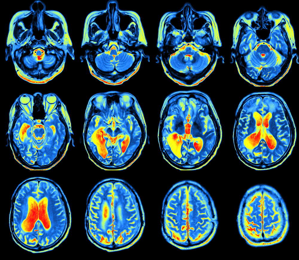
All participants were evaluated by Voxel-Based Morphometry (VBM) to determine the brain anatomy, diffusion-tensor imaging (DTI) for the analysis of brain area connectivity, and functional MRI (fMRI) to assess brain activity. Patients also underwent a complete cognitive and clinical assessment.
The MRI-VBM analysis revealed that Friedreich’s ataxia patients had some grey matter areas (the more superficial layer) with reduced volume compared to healthy volunteers. Specifically, the areas were in the lower region of the brain, just above the cerebellum, which suggests “an alternative sensorimotor cerebellar map involvement in Friedreich’s ataxia,” the researchers wrote.
An analysis of the white matter (the tissue that goes deeper into the brain) by MRI-DTI also revealed the existence of connectivity alterations. The most affected areas were the inferior and superior cerebellar peduncles, which are responsible for transmitting information from the cerebellum to other parts of the central nervous system.
Evaluation of the brain activity did not show any differences as to what concerned the control movement of the dominant hand, whereas to control of the nondominant hand, the brain responded differently in each group.
This particular difference had not been reported in any previous studies, probably because this type of motor test is often applied only to the dominant hand and not the other, the researchers said.
Combined with MRI-DTI data, this finding suggests that cerebral-cerebellar circuitry disruption may affect both hemispheres.
Overall, the brain reactivity as determined by functional MRI was found to correlate with the clinical parameters of Friedreich’s ataxia.
And evaluation of the IQ level was found to be within normal range in 13 patients, while four patients were considered to be borderline, and two had intellectual disabilities. In four of these patients with reduced IQ, the verbal performance was more impaired.
The researchers did not find any significant link between DTI and functional MRI measurements and IQ values.
Collectively, these findings “bring a new dimensional role of the cortical circuitry [the outer sheet of the brain] involved in Friedreich’s ataxia,” the researchers stated.


Leave a comment
Fill in the required fields to post. Your email address will not be published.