Atrophy Along Spine and in Specific Brain Areas Linked to Clinical Decline in FA Patients, Study Reports


Patients with Friedreich’s ataxia (FA) have smaller spinal cords and evidence of atrophy in specific areas of the brain — differences that, in their degree, work to help predict clinical decline, a German study reports.
The study, “Structural characteristics of the central nervous system in Friedreich ataxia: an in vivo spinal cord and brain MRI study,” appeared in the Journal of Neurology, Neurosurgery & Psychiatry.
Prior research using magnetic resonance imaging (MRI) found that spinal cord atrophy correlates with disability in FA. But these studies of spinal alterations, including reduced area, focused on upper cervical areas and not the full spinal cord.
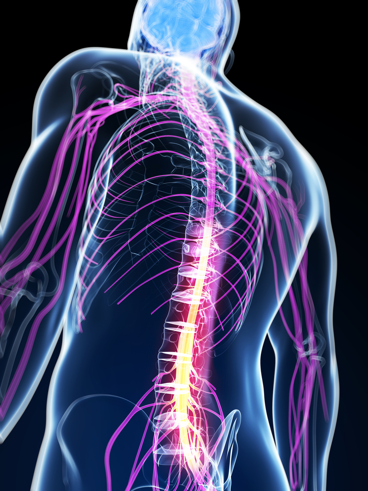
Researchers at several German institutions studied both the cervical and thoracic spinal cord in FA patients. While the cervical spine contains the first seven vertebrae, mainly located in the neck, the thoracic spine is composed of 12 vertebrae and located in the mid-back, where it holds the rib cage and protects the heart and lungs.
Besides these spinal measurements, they also did MRI scans of the brain. Degeneration of the cerebellum — which coordinates balance and movement — and abnormal structure in other brain areas are known in FA.
Overall, the research team assessed whether spinal and brain alterations could predict disease severity. To do this, they compared 21 patients (mean age 35, 10 males) with 22 healthy controls matched for age and sex. They also compared 14 patients with severe gait impairment with seven who were less impaired.
Besides spinal cord and brain MRI scans, appropriate scales were used to assess ataxia — the lack of coordination in voluntary movements – non-ataxia signs, like spasticity and resting tremor, and patients’ ability to conduct activities of daily living (ADL). Examinations of motor, speech and other skills, using the Spinocerebellar Ataxia Functional Index (SCAFI) timed tests, were also conducted.
Results revealed that, in comparison with controls, the entire spinal cord of FA patients was reduced, particularly the cross-sectional area of the cervical cord. The thoracic cord, and to a lesser extent the cervical cord, also showed pronounced flattening or compression.
Data further showed that patients with severely gait difficulties had reduced mid-thoracic cross-sectional area and volume than did those with lesser impairment.
In terms of brain volume, the most pronounced atrophy was found in areas called medulla oblongata — the lower part of the brainstem, responsible for involuntary functions such as breathing or blood pressure; the pons – also involved in involuntary functions, as well as facial expressions; and the midbrain, which regulates processes that include motor control, hearing, vision, and sleep.
The researchers also observed reduced volumes in the cerebellum and the diencephalon, which is surrounded by the cerebral hemispheres and includes the thalamus and the hypothalamus.
Subsequent analyses showed that shrinkage (reduced volume) in the cervical and thoracic areas of the spinal cord were associated with genetics, age at FA onset, disease duration and clinical scores. Poorer volume in the lower brainstem and cerebellar area correlated with clinical impairment.
Further links were found between changes in specific parts of the cerebellum and ratings of ataxia and ADL, as well as between alterations in the white matter — made of nerve fibers connecting grey matter areas — of the cerebellum and both non-ataxia signs and SCAFI scores.
Combining measures of “both brain and spinal cord size reductions may yield better prediction of disease progression in [FA],” the researchers added, noting that such clinical markers of disease-relevant changes over time may be of particular importance to clinical research into FA.