#IARC2017 – Mitochondria, Deprived of Frataxin Protein, Fail to Control Free Radicals in Cells, FA Study Finds
Written by |


In Friedreich’s ataxia, insufficient frataxin protein in cells impairs the ability of mitochondria to control the production of harmful reactive oxygen species, which accumulates and ultimately triggers nerve cell death, research into a mouse model of the disease revealed.
The study, detailing how harm done to mitochondria in nerve cells helps to promote disease progression, was conducted by researchers at the University College London (UCL) Institute of Neurology, and presented today by Rosella Abeti at IARC 2017, the International Ataxia Research Conference taking place at Pisa, Italy, through Sept. 30.
Her presentation was titled “Addressing mitochondrial function in a mouse model of Friedreich’s Ataxia (FRDA).”
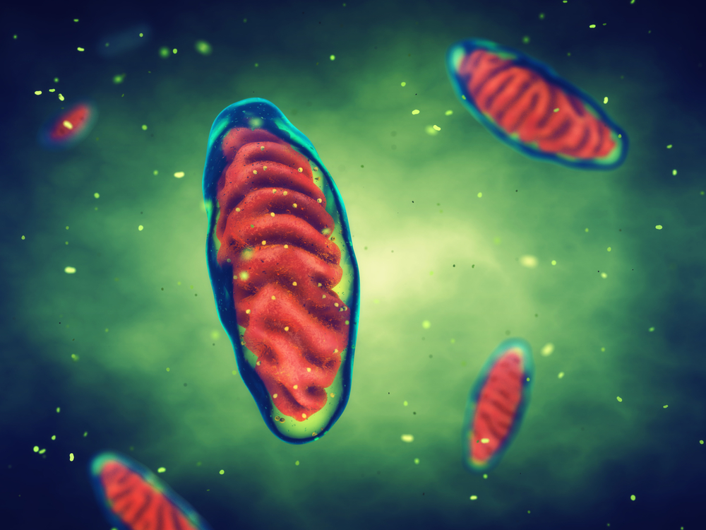
Frataxin is a protein found in mitochondria, the small organelles inside cells where energy is produced. Mutations in the frataxin gene — specifically a GAA repeat expansion — leads to low levels of frataxin protein production in people with Friedreich’s ataxia (FA), which changes how mitochondria function.
Several studies over the past two decades, working with Friedreich’s ataxia patients and mouse models of the disease, have investigated its underlying mechanisms. But the role of frataxin in mitochondrial health is still poorly understood.
This study investigated how low levels of frataxin impact mitochondria. Researchers used live cell microscopy and assessed several parameters of mitochondria’s function using nerve cells (neurons) of the YG8R Friedreich’s ataxia mouse model.
Mice were depleted of their gene for frataxin and genetically engineered to produced the frataxin human mutated gene, carrying several GAA repeat expansions.
Mitochondrial function was assessed by monitoring the decline in mitochondrial membrane potential, which triggers cells death, and by monitoring the maintenance of mitochondria redox homeostasis, or the ability to maintain an environment free of oxidative stress. Stressful cellular environments are characterized by the production of harmful reactive oxygen species (ROS, or free radicals), unstable oxygen-containing molecules that react easily with other molecules in cells, damaging and potentially killing them.
Results showed that mitochondria in Friedreich’s ataxia-like neurons are dysfunctional, and that a reduction in frataxin levels resulted in a decrease of mitochondrial membrane potential.
This imbalance led to oxidative stress and the generation of damaging and highly reactive ROS. In turn, ROS accumulation inside neurons led to lipid damage — a process known as lipid peroxidation — which, if allowed to persist, leads to cell death. Lipids are the major constituents of cellular membranes.
Preventing an increase in lipid peroxidation in neurons protected against cell death, the researchers showed. The team used three drugs to prevent lipid peroxidation: PUFA, SFN, and RTA 408 (omaveloxolone).
In her presentation, Abeti detailed the changes seen in mitochondria function in neurons from a Friedreich’s ataxia mouse model, and suggested that lipid peroxidation could be an important treatment target.