Friedreich’s Ataxia Study Sheds Light on Molecular Development of Disease

The mechanism leading to cell death in Friedreich’s ataxia has been described in a new study published in the Annals of Clinical and Translational Neurology.
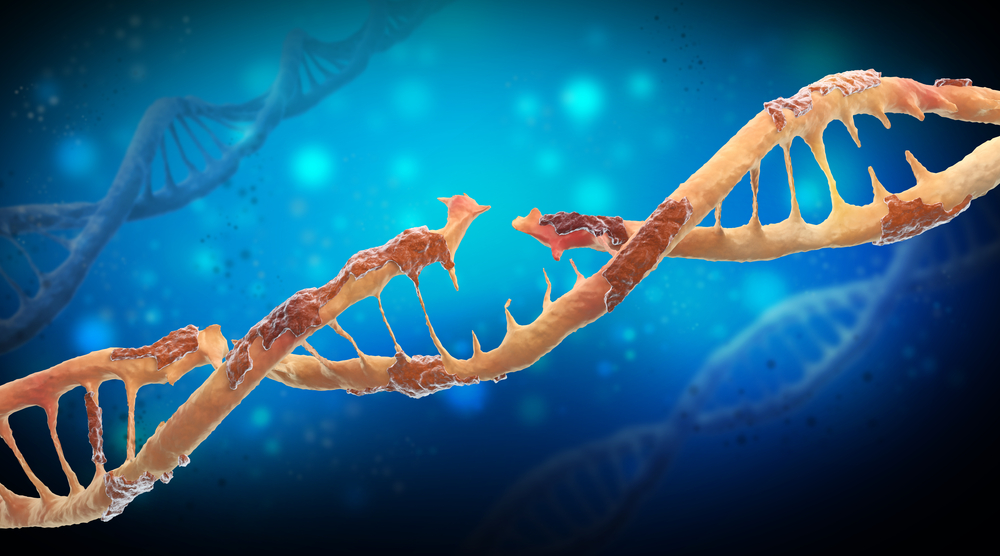
According to the study, “Deep sequencing of mitochondrial genomes reveals increased mutation load in Friedreich’s ataxia,” down-regulation of a gene called frataxin (FXN) increases damage to mitochondrial DNA (mtDNA). This increase in the mutation load of the mitochondrial genome reduces DNA repair capacity. With time, mtDNA mutations accumulate and reduce mitochondrial fitness, leading to cell death and the symptoms of Friedreich’s ataxia (FA).
The team of researchers, led by Dr. Marek Napierala of the University of Alabama in Birmingham, measured the amount of mtDNA damage and mutation load in Friedreich’s ataxia and control fibroblasts grown in the laboratory. They also analyzed the ability of Friedreich’s ataxia and control cells to repair oxidative damage in their mtDNA and the status of the genes involved in DNA repair and metabolism.
They saw that short or long-term down regulation of FXN gene expression results in a significant increase in mtDNA damage, which translates in a significant increase in mutation load in mtDNA. Low FXN gene expression also reduces the ability of the cells to repair mtDNA damage caused by oxidative stress.
The results revealed that Friedreich’s ataxia fibroblasts had a higher level of mtDNA damage compared to unaffected cells, and that control cells (depleted of FXN) acquire mtDNA damage in a manner similar to cells in Friedreich’s ataxia patients.
“We propose that accumulation of mtDNA lesions represents a critical event in the development and progression of [Friedreich’s ataxia],” the authors wrote. “Next‐generation sequencing of Friedreich’s ataxia mitochondrial genomes revealed a widespread increase in mutation load in patient fibroblasts. Although mtDNA damage alone can have profound consequences within the cell, low expression of FXN may also affect the nuclear genome.”
“Therefore, it would be of interest to extend these studies into whole‐genome analyses of Friedreich’s ataxia samples including terminally differentiated neurons, glia, and cardiac cells,” the authors concluded.


