Nerve Fiber Growth, Repair Inhibited in Friedreich’s Ataxia, Study Finds
Written by |

Mutations that cause Friedreich’s ataxia lead to abnormalities in how nerve fibers grow and reduce their ability to repair themselves, according to a study done in mouse cells.
These differences may contribute to nerve cell degeneration that is characteristic of the disease, researchers say.
The study, “Frataxin Deficit Leads to Reduced Dynamics of Growth Cones in Dorsal Root Ganglia Neurons of Friedreich’s Ataxia YG8sR Model: A Multilinear Algebra Approach,” was published in the journal Frontiers in Molecular Neuroscience.
Friedreich’s ataxia is caused by mutations in the gene encoding frataxin, an important mitochondrial protein. The specific mechanisms of how these mutations cause abnormalities in various parts of the body are still being worked out. For example, patients diagnosed with Friedriech’s ataxia characteristically experience degeneration in the dorsal root ganglia (DRG), a group of cell bodies responsible for transmitting sensory messages and located on spinal roots, but the reasons for this degeneration are largely unknown.
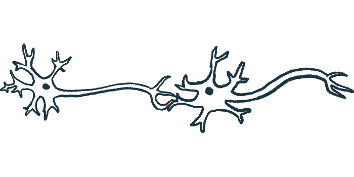
Neurons (nerve cells) have long, wire-like projections called axons, which the cell uses to connect with other nerves or other parts of the body. When a new axon is developing or a damaged one is being repaired, a structure called a growth cone, or GC, is formed at the tip of the budding axon. This structure basically builds out the axon until it connects with its intended target.
In this study, an international team of scientists recorded videos and images of GC growth for DRG neurons from mice — either a model of Friedreich’s ataxia called YG8sR or control mice. The team then used sophisticated mathematical analyses to examine differences in the dynamics of GC growth over time.
A number of abnormalities were found in the GCs from YG8sR neurons (YG8sR-GCs). Compared to control GCs, those with disease-causing mutations moved significantly slower and covered less overall distance; they also exhibited abnormalities in how they turned to redirect growth. Differences in the shapes of YG8sR-GCs also were noted, though only in younger mice.
“Overall, the changes in the spatial behavior of YG8sR-GCs suggest that frataxin deficient YG8sR neurons can build GCs, but these would be dysfunctional,” the researchers concluded.
Since growth-cone function is important for repairing damaged axons, the scientists next conducted cell experiments testing the neurons’ ability to regenerate axons after injury. Results showed that axon repair was significantly impaired in YG8sR neurons.
“Our results show that while control DRG neurons can grow healthy axons with well-defined tracks in response to a [repair-promoting chemical signal], the YG8sR-DRG neurons cannot do it,” the scientists wrote.
“Frataxin deficient DRG neurons from the YG8sR mice may lose the intrinsic ability to regenerate their axons after injury, a protective phenomenon observed mainly in DRG neurons,” they added. “A possible explanation for this observation is the failure of axonal regeneration programs in the YG8sR-DRG neurons, which in normal conditions allows them to protect against cumulative axon damage.”
The scientists speculated that these alterations in growth cone dynamics and axon repair may contribute to degeneration in the dorsal root ganglia, though they stressed that “further analysis of the GC dynamics and axonal regeneration response is needed to reveal the cascade of molecular events that leads to the dysfunction of the DRG-GC in” Friedriech’s ataxia.


