
Functional MRI Used to Study of Neuropathology of Spinocerebellar Ataxia Type 7
Written by |


 A post-doctoral fellowship award has been given to Carlos Roberto Hernandez-Castillo, Ph.D. of Universidad Nacional Autonoma de Mexica, Coyoacan, Mexico to study the neuropathology of spinocerebellar ataxia type 7 using functional magnetic resonance imaging (fMRI).
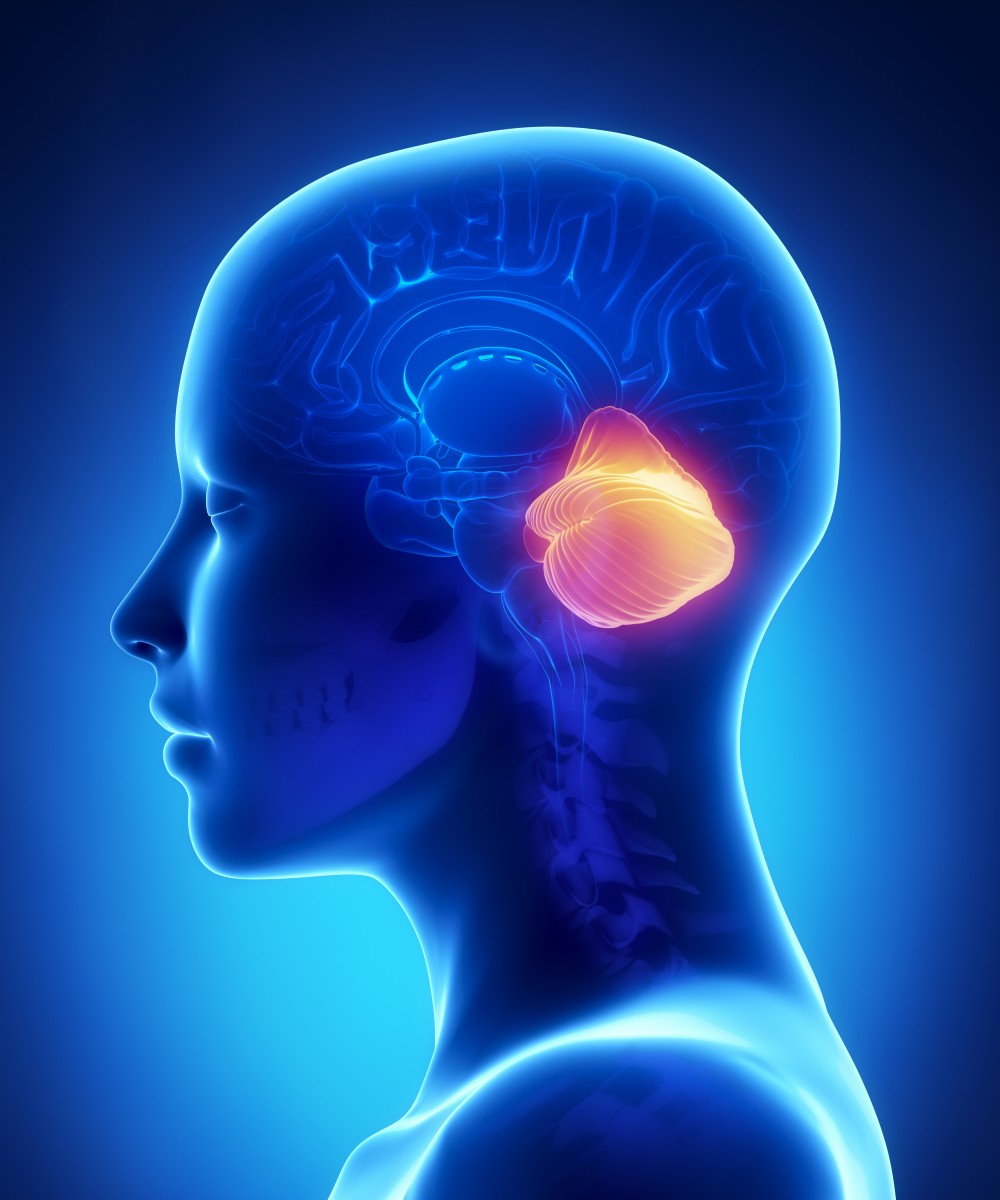
Spinocerebellar ataxia type 7 (SCA7) is an extremely rare genetic disorder that affects a brain area known as the cerebellum, which controls coordination. It is caused by a repeated region of the SCA7 gene. An effective treatment or cure is not available for SCA7, which can affect anyone of any age.
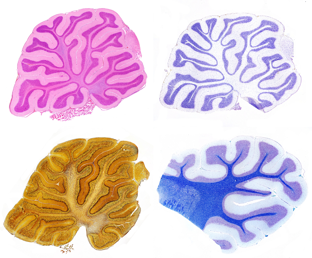
Previous research has shown that specific areas of the cerebellum die off in SCA7, called the olivopontocerebellar regions. In addition, the retina can also degenerate.
Hernandez-Castillo has already studied this and many other changes that occur in the disease, using fMRI, stating that “Recent imaging studies carried out in our lab have expanded the knowledge of the pathological changes resulting from the SCA7 degenerative process, including white matter reduction, grey matter atrophy and functional connectivity abnormalities. However, all those reports have been performed in cross-sectional studies.” In a cross-sectional study, data is only collected at one specific time. To really understand SCA7, data needs to be collected over multiple time-points, so see how the disease progresses.
The newly funded study will focus on SCA7 patients over 2 years. Twenty-six patients that have been confirmed as having SCA7 using genetic tests will participate in this study. The researchers will use fMRI to take images of the brain over time. They will also measure movement problems in the study subjects, as well as problems with cognition and vision. According to a description of the study, “The proposed analysis might help understand what are the most early observable changes that may be useful as biomarkers to be used along specific therapies or treatments in the early stages of the disease.”
Changes in the fMRI images may serve as biomarkers, which are measurable biological changes that can indicate a disease or condition. In this case, the biomarkers would consist of a pattern of brain activity, as measured by fMRI.
This was one of the 23 research studies from the United States, Belgium, Mexico, United Kingdom, Portugal, and Germany recently funded by The National Ataxia Foundation (NAF). According to their mission statement the NAF is “dedicated to improving the lives of persons affected by ataxia through support, education, and research.” With the funding of these and previous research studies granted research money earlier in 2014, nearly one million dollars was awarded for ataxia research by the NAF.


Leave a comment
Fill in the required fields to post. Your email address will not be published.