
Brain Structure Abnormalities in Late-onset Friedreich’s Ataxia Not Linked to Disease Features
Written by |


People with late-onset Friedreich’s ataxia have similar, but not identical, abnormalities of brain structures as those who become ill at an earlier age, researchers recently demonstrated. The study sheds light on why the features of the disease differ in people with early and late onset.
Researchers also argued that their findings — presented in the study, “Structural Signature of Classical Versus Late-Onset Friedreich’s Ataxia by Multimodality Brain MRI” — might add knowledge to the search for imaging biomarkers of disease in Friedreich’s ataxia. The work was published in the journal Human Brain Mapping.
To compare the brains of patients with early and late-onset disease, researchers at the School of Medical Sciences of the University of Campinas in Brazil recruited 36 Friedreich’s ataxia patients. Among them, 13 had developed the condition after the age of 25, and were considered late-onset patients. The research team also recruited 29 healthy volunteers to use as a comparison.
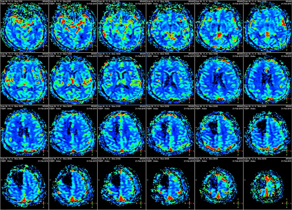
Using magnetic resonance imaging (MRI) brain scans, the team assessed both gray and white matter parts of the brain. The gray matter is composed mainly of nerve cell bodies, while white matter is made up of long neuron appendages connecting different parts of the brain.
Both early- and late-onset Friedreich’s ataxia patients had areas in their gray matter that had an abnormally small volume. The brain’s motor cortex was among the areas apparently most affected by the disease. This superficial brain area mainly deals with movement and showed a reduced thickness in both patient groups, compared to controls.
Both groups also had abnormal structures in several white matter regions. They noted subtle differences between patients who became ill at an older age and those with early-onset disease in several brain regions.
Patients who developed ataxia at an earlier age had more widespread abnormalities of a sort researchers refer to as microstructural.
When the research team attempted to correlate brain imaging findings to symptoms, they found that gray matter volume reductions in certain areas corresponded to disease duration and severity in early-onset patients. The brain abnormalities in late-onset Friedreich’s ataxia patients could not be linked to any disease features. Neither could abnormal structures of white matter regions be linked to any disease aspects.
“These results provide meaningful insights into disease biology and also add relevant information on the use of neuroimaging metrics as biomarkers for [Friedreich’s ataxia]. The multiatlas approach proved to be a useful tool to identify biomarkers in [Friedreich’s ataxia], which might help upcoming clinical trials,” the authors concluded.


Leave a comment
Fill in the required fields to post. Your email address will not be published.