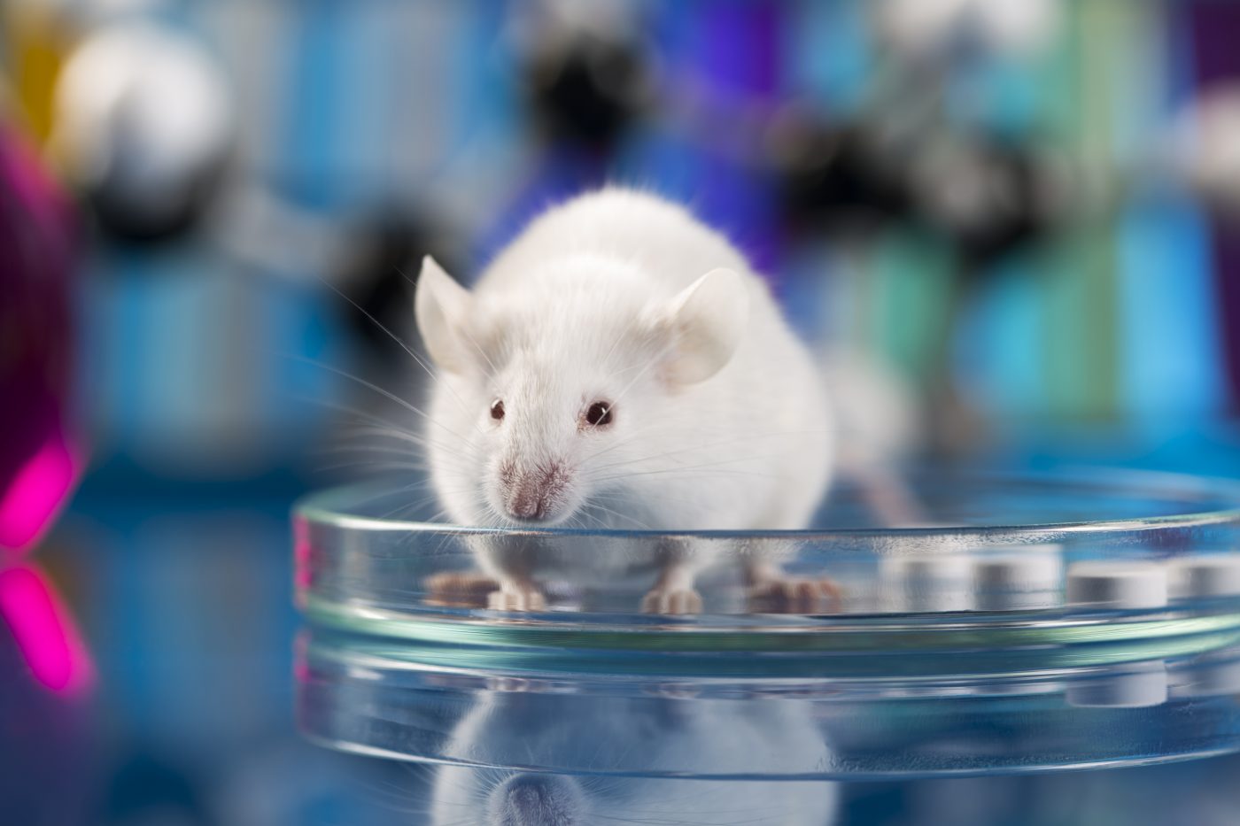
FA Mouse Models May Not Fully Mimic Human Disease

Truncated forms of frataxin, the protein that is lost in people with Friedreich’s ataxia (FA), were found predominantly in mouse tissues, contrasting with the dominance of non-truncated frataxin in human tissue, a study found.
The findings have implications for mouse models used to mimic the condition in disease research and therapeutic development, the scientists said.
The study, “Extra-mitochondrial mouse frataxin and its implications for mouse models of Friedreich’s ataxia,” was published in the journal Nature Scientific Reports.
FA is caused by a deficiency in the protein frataxin, which supports the function of enzymes within the mitochondria, the small structures in cells that produce energy. Frataxin is found throughout the body, but is highest in the heart, spinal cord, liver, pancreas, and skeletal muscles used for movement.
Many frataxin-deficient mouse models have been developed to understand the underlying disease mechanisms and evaluate potential therapies. In these models, the specific form of mouse frataxin protein is assumed to be the same as human frataxin.
In mice, full-length frataxin is produced in the cell as a protein composed of 207 amino acids — the building blocks of proteins. This full-length protein is transported into the mitochondria and cleaved in two steps into a smaller functional (mature) form with 130 amino acids, analogous to the 130-amino acid human frataxin.
However, recent studies suggest there are multiple forms of mouse frataxin. Furthermore, an alternate form of human frataxin was discovered outside the mitochondria, raising the possibility of a corresponding form of mouse frataxin outside the mitochondria.
Given these new findings, further investigation is needed to determine if multiple forms of frataxin exist, which may uncover unknown roles of frataxin outside the mitochondria in FA and support the development of more accurate FA mouse models.
Now, scientists at the University of Pennsylvania have isolated and characterized major forms of frataxin in mouse heart, brain, and liver, as well as human heart tissue, using an antibody that specifically identifies and binds to both human and mouse frataxin.
The analysis revealed three shortened (truncated) forms of frataxin in mouse heart and brain tissue, a predominant form missing one amino acid, making it 129-amino acids, and two minor forms of 127- and 124-amino acids. Two primary frataxin forms were found in liver tissue, 129- and 127-amino acids, and three minor forms of 128-, 125- and 124-amino acids.
The predominant truncated form of frataxin (129-amino acids) accounted for 77.2% of mature frataxin present in the mouse heart, 86.9% in the brain, and 47.0% in the liver. The longer 130-amino acid mouse frataxin, matching the mature human protein, represented only 14.9% of mature frataxin in the heart, 7.3% in the brain, and 11.3% in liver tissue.
The second major 127-amino acid protein occurred in 36% of liver tissue, which was “barely detected in mouse heart and was not detected in mouse brain indicating there might be an additional role for this [form] in mouse liver,” the team wrote.
A closer examination of mouse liver cells found that the major frataxin protein observed in the cell cytosol — outside the nucleus and mitochondria — was the 129-amino acid form, accounting for 96% of the protein.
The remaining 4% was the predicted 130-amino acid mitochondrial form of the protein, but researchers believe this form “most likely arose in the cytosol through contamination by disrupted mitochondria during freeze thawing,” they wrote.
Full-length frataxin (130-amino acids) and intermediate frataxin (167-amino acids) from the first cleavage step were not detected in the cytosol of mouse liver cells. The mature 130-amino acid frataxin was more enriched in the mitochondria (36.4%) and the nucleus (29.8%) compared to the cytosol.
The presence of an elongated form of frataxin, previously found outside mitochondria in human cells, was not identified in mouse heart, brain, or liver tissue.
Finally, an analysis of postmortem heart tissue isolated from five people with no history of heart failure found the concentration of mature human frataxin (130-amino acids) was 7.9 nanograms per mg (ng/mg), which was similar to the total frataxin forms in mouse heart tissue of 5.5 ng/mg.
In contrast to mouse frataxin, the major form of human frataxin in each of the five human heart samples was the mature 130-amino acid form, corresponding to 98.4% of the mature frataxin. Low amounts of a 129-amino acid form (0.1%) and a 128-amino acid frataxin (1.5%) were observed, showing that “frataxin in human heart was present primarily as mature frataxin,” the team wrote.
“The major frataxin [form] in the mouse is the 129-amino acid truncated mature frataxin and not the 130-amino acid form of mature frataxin,” the investigators concluded, adding that the “processing of the mature mouse frataxin could proceed in a different manner than in humans.”
“If this is the case, we would suggest that mouse models do not serve as a good model for humans,” they added.


