Low Levels of Friedreich’s Ataxia-linked Protein Don’t Impact Red Blood Cells, Study Suggests
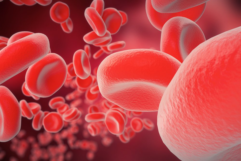


Low levels of frataxin protein, the hallmark of Friedreich’s ataxia, do not seem to impact the function of red blood cells, particularly the formation of hemoglobin’s core molecule, heme, a study suggests.
The finding casts doubt on some researchers’ supposition that iron-metabolism problems play a role in Friedreich’s ataxia. It also explains why anemia — lack of iron stemming from impaired heme production — is not a problem in Friedreich’s ataxia patients.
Reseachers published their study, “No changes in heme synthesis in human Friedreich´s ataxia erythroid progenitor cells,” in the journal Gene.
Friedreich’s ataxia, which affects 1 in 50,000 people, is the most common form of inherited ataxia, a neurodegenerative disease. Low levels of frataxin trigger the disease, but the protein’s precise role in the process is still unclear.
Some studies have suggested that frataxin could play a role in iron metabolism, particularly metabolism mediated by cell components called mitochondria. Scientists refer to mitochondria as the cells’ powerhouses because of their key role in energy production.
One reason some researchers have speculated that frataxin plays a part in iron metabolism is that the protein is capable of binding to iron. Another is that iron metabolism is deregulated in Friedreich’s ataxia.
Heme is a crucial component of hemoglobin, the protein in our red blood cells that carries oxygen throughout the body. Because heme is formed partly in mitochondria, researchers decided to investigate whether frataxin played a role in iron metabolism and heme formation.
They analyzed erythroid progenitor cells — self-renewing stem cells that give rise to red blood cells — from three Friedreich’s ataxia patients and three healthy controls. Progenitor cells were chosen because they are a long-established model for heme studies.
The team measured several biomarkers of heme formation, including intracellular iron levels and hemoglobin formation, in cells from the Friedreich’s ataxia patients and the controls.
As expected, they found significantly less frataxin in the Friedreich’s ataxia cells then in the controls’ cells. They also found no significant differences between the groups in heme formation parameters such as hemoglobin and iron.
This gibed with the fact that most Friedreich’s ataxia patients do not have anemia. The impaired heme formation that causes anemia leads to a drop in red blood cells as well.
The team also compared gene expression in cells from Friedreich’s ataxia patients and controls. They discovered differences in the expression of 11 genes linked to oxidative stress, an imbalance between the body’s production of free radicals and its ability to counter the radicals’ harmful effects. Research has linked oxidative stress to other neurodegenerative diseases, such as Alzheimer’s and Parkinson’s.
Notably, the team that did the Friedreich’s ataxia study found no differences in iron parameters between the patients and controls.
Overall, the results “indicate that the main regulatory network of frataxin is rather involved in oxidative stress than in iron metabolism,” the researchers wrote.