Gene Mutation in Friedreich’s Ataxia Fruit Flies Caused Iron Toxicity, Death of Brain Cells


The frataxin gene regulates iron storage in cells, but mutations in the gene are known to cause Friedreich’s ataxia (FA). A study now shows the mutation causes iron toxicity and the loss of nerve cells in fruit flies, an effect prevented when they were put on a low-iron diet.
The study, “Loss of Frataxin induces iron toxicity, sphingolipid synthesis, and Pdk1/Mef2 activation, leading to neurodegeneration,” published in eLIFE, demonstrates the first evidence showing how mutations in the fraxatin gene could lead to the progressive death of brain cells observed in Friedreich’s ataxia patients.
In FA, nerve cells in the spinal cord and some parts of the brain progressively lose their functions and die upon aging (neurodegeneration). This results in difficulties in walking, along other conditions such as heart disease and diabetes.
Researchers have known a mutation in the frataxin gene is the primary cause behind FA. The gene encodes a protein normally found in mitochondria – the energy-producing powerhouses inside cells. Previous studies suggest that mutations in frataxin prevent mitochondria from working normally, which triggers the production of toxic chemicals called reactive oxygen species (ROS).
The frataxin protein has also been proposed to act as an iron storage protein and as such, subsequent mutations lead to an accumulation of toxic iron levels within cells. But it is still a matter of debate whether the accumulation of iron contributes to Friedreic’s ataxia, and the mechanisms behind cell death are still unknown.
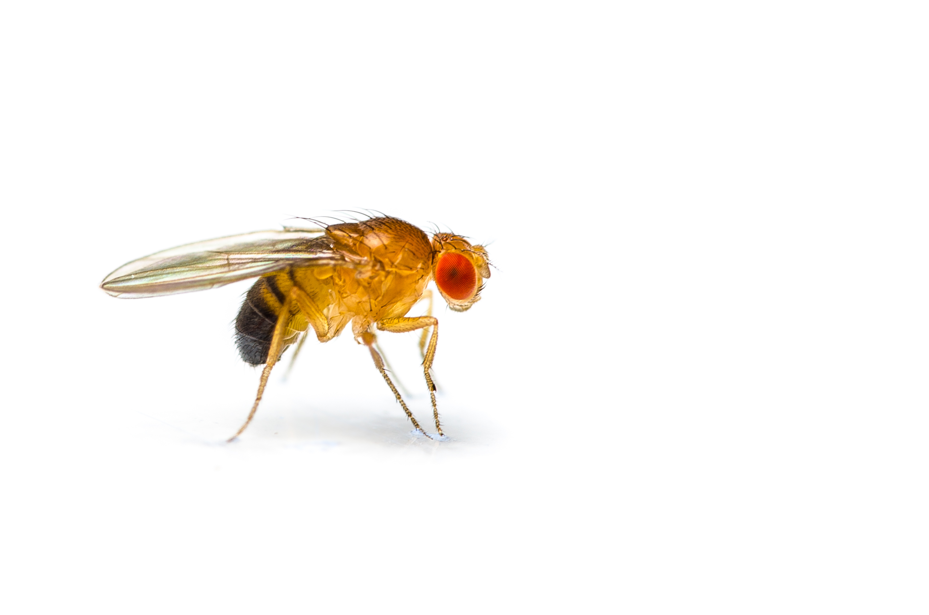
Researchers wanted to identify other mechanisms that could explain the loss of nerve cells seen in Friedreich’s ataxia using fruit flies as an experimental model. They investigated iron levels and neurodegeneration (gradual death of brain cells) in fruit flies lacking the fraxatin gene.
They found that the loss of the gene caused iron to increase in brain cells and other tissues and was followed by enhanced neurodegeneration, but did not produce more reactive oxygen species. The team also identified a novel molecular pathway that explains how iron toxicity causes cellular death: Iron buildup increased the production of fatty molecules (called sphingolipids), which in turn triggered the activation of two proteins (Pdk1 and Mef2).
Blocking any of these effects could delay cellular death in the fruit fly models and neurodegeneration was prevented when the flies were served a low-iron diet.
As with many degenerative diseases of the nervous system, there is currently no cure or effective treatment for Friedreich’s ataxia. Therapies are currently focused on the easing the symptoms and accompanying complications that can be treated to help people maintain optimal functioning as long as possible.
Researchers said additional studies are needed to see if these effects hold true in animal and human studies. Their findings provide potential new targets for future therapies for Friedreich’s ataxia.