NAF Supports Study Exploring Mitochondrial Defects in Skeletal Muscle in Cerebellar Ataxia 2


The National Ataxia Foundation (NAF) recently awarded four post-doctoral fellowship grants covering various aspects of ataxia research. In a project focusing on autosomal recessive cerebellar ataxia 2, Pankaj Kumar Singh at the Institut Génétique Biologie Moléculaire Cellulaire, in France, explores how mitochondrial defects contribute to disease mechanisms in skeletal muscle, hoping to identify a therapeutic target in type 2 cerebellar ataxia.
Dr. Singh is one of four researchers awarded the grant, and his work will center on a mouse lacking Adck3, the gene underlying this type of ataxia. The mouse, recently created in Dr. Singh’s lab, exhibits many of the features of cerebellar ataxia 2, including ataxia and mild exercise intolerance. These mice also have a mitochondrial defect that is particularly evident in skeletal muscle.
Based on similarities to a protein in yeast, the researchers believe that the Adck3 protein is a factor in mitochondria, playing a role in the synthesis of Coenzyme-Q, but little is known about the molecular mechanisms of this protein in cerebellar ataxia 2.
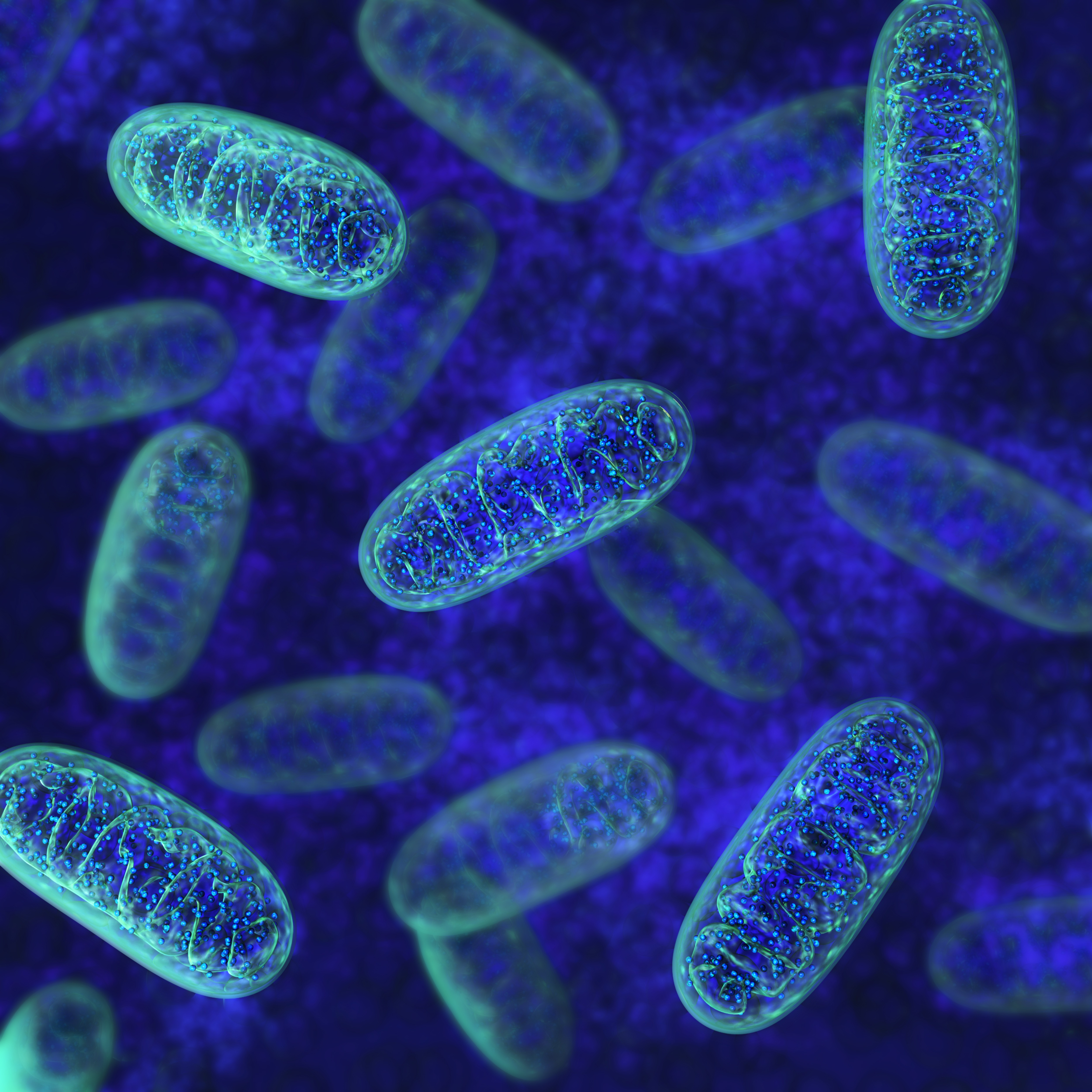
Mitochondria are key energy producers in the body, converting nutrients to energy using a series of complex oxidative events. Because of their crucial role for metabolism, dysfunctional mitochondria are linked to a range of diseases. One of the consequences of the inability of mitochondria to perform their work is a reduced oxygen consumption, paired with an increased anaerobic metabolism, leading to exercise intolerance — a feature of cerebellar ataxia 2.
Dr. Singh hypothesizes that the exercise intolerance seen both in patients and in the Adck3-deficient mice is a consequence of mitochondrial dysfunction in skeletal muscle.
The project has the ambitious goal of identifying a molecular target — linked to the mitochondrial deficit in skeletal muscle — that can be manipulated to relieve symptoms and improve exercise performance in the mouse model. Such findings would be a promising contribution to potential drug development attempts in type 2 cerebellar ataxia.
Dr. Singh’s project also intends to explore molecular pathways in the cerebellum affected by the lack of Adck3 in these mice. Such pathways are likely linked to the cerebellar dysfunction in ARCA2, and their identification might advance the current understanding of this type of ataxia.