
#IARC2017 – Small Molecules Mimicking Brain Factors Raise Frataxin Protein Levels in FA Mice in Study
Written by |


Administering small molecules that mimic factors naturally found in the brain to a mouse model of Friedreich’s ataxia increased levels of frataxin expression in both the brain and heart — two organs highly affected in people with the disease.
The study, “Neurotrophic factor and cytokine mimetics as new potential therapeutic agents for Friedreich’s ataxia,” was presented by Javier Díaz-Nido with the Centro de Biologia Molecular Severo Ochoa, Universidad Autonoma de Madrid, Spain, on Saturday, the final day of IARC 2017, the International Ataxia Research Conference held in Pisa, Italy.
The presentation was part of Session 4 of the meeting, looking at “Therapeutics and Clinical Trials.”
Low frataxin protein levels, a result of mutations in the frataxin gene, is the underlying cause of Friedreich’s ataxia (FA), a neurodegenerative disease. Degeneration in the spinal cord and peripheral nerves, and to a lesser extent in the cerebellum (the brain region that coordinates balance and movement), leads to unsteady movements and impaired sensory functions.
Previous studies suggest that neurotrophic factors like the Brain-Derived Neurotrophic Factor (BDNF) and cytokines like Erythropoietin (Epo) could prevent the neurodegeneration triggered by low levels of frataxin. They also were seen to increase frataxin protein levels in different experimental models.
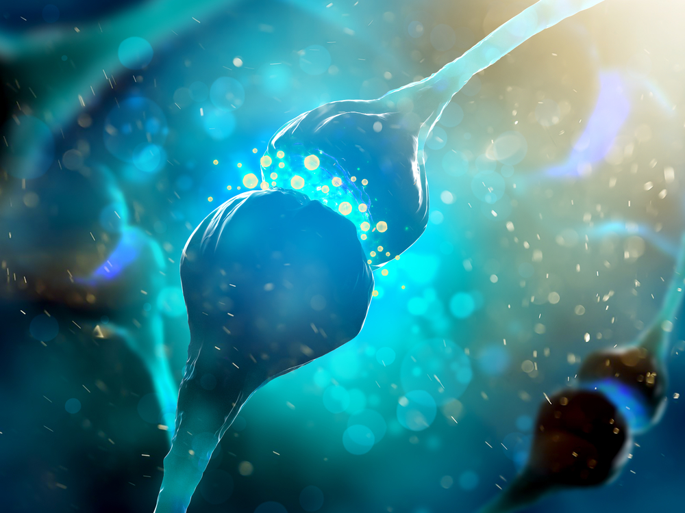
Neurotrophic factors are a family of molecules that support the growth, survival, and differentiation of nerve cells (neurons). Cytokines are small secreted proteins released by cells, and they work as chemical messengers being capable of influencing the behavior of other cells.
Despite their potential benefits, therapeutic use of these molecules has been limited due to their poor pharmacokinetic properties — how a drug behaves once in the body — and their inability to effectively cross into the brain by passing through the blood-brain barrier.
“In order to address these limitations, we have explored the therapeutic potential of smaller molecules which can activate BDNF or Epo receptors,” the researchers wrote.
One of these molecules, called 7,8-dyhydroxyflavone (7,8-DHF), activates the BDNF receptor, thereby mimicking the effects of BDNF itself.
Results showed that 7,8-DHF added in vitro to primary neuron cell cultures — taken from healthy mice serving as controls and from YG8 mice, a mouse model of FA — both decreased the expression of cell death markers and increased levels of the frataxin protein.
This increase was also detected when 7,8-DHF was added to stem cells derived from FA patients.
Researchers then tested the molecule in animal models of Friedreich’s ataxia, and saw a response in the cerebellum but not the heart.
“Interestingly, the systemic administration of 7,8-DHF to YG8 mice in vivo enhances frataxin expression in the cerebellum but not in the heart of treated animals,” they wrote. This finding, actually, is in agreement with the fact that there is a higher expression of BDNF receptors in the brain.
A molecule that activates Epo receptors — the helix B surface peptide (HBSP) — was also seen to increase frataxin expression in primary neuron cell cultures from both normal and YG8 mice, and from FA patient-derived stem cells.
Furthermore, systemic administration — a treatment that circulates in the bloodstream — of HBSP to YG8 mice increased frataxin levels in both the cerebellum and the heart.
“In view of these data we suggest that neurotrophic factor and cytokine mimetics such as 7,8-DHF and HBSP should be considered as a novel strategy to increase frataxin expression and delay neurodegeneration in FRDA [Friedreich’s ataxia],” the researchers concluded.


Leave a comment
Fill in the required fields to post. Your email address will not be published.