Friedreich’s Ataxia Patients Have Altered Connectivity Between Brain Regions, Study Shows
Written by |


People with Friedreich’s ataxia (FA) have alterations in the connectivity of different brain regions, which most likely is associated with the disease’s neurological symptoms.
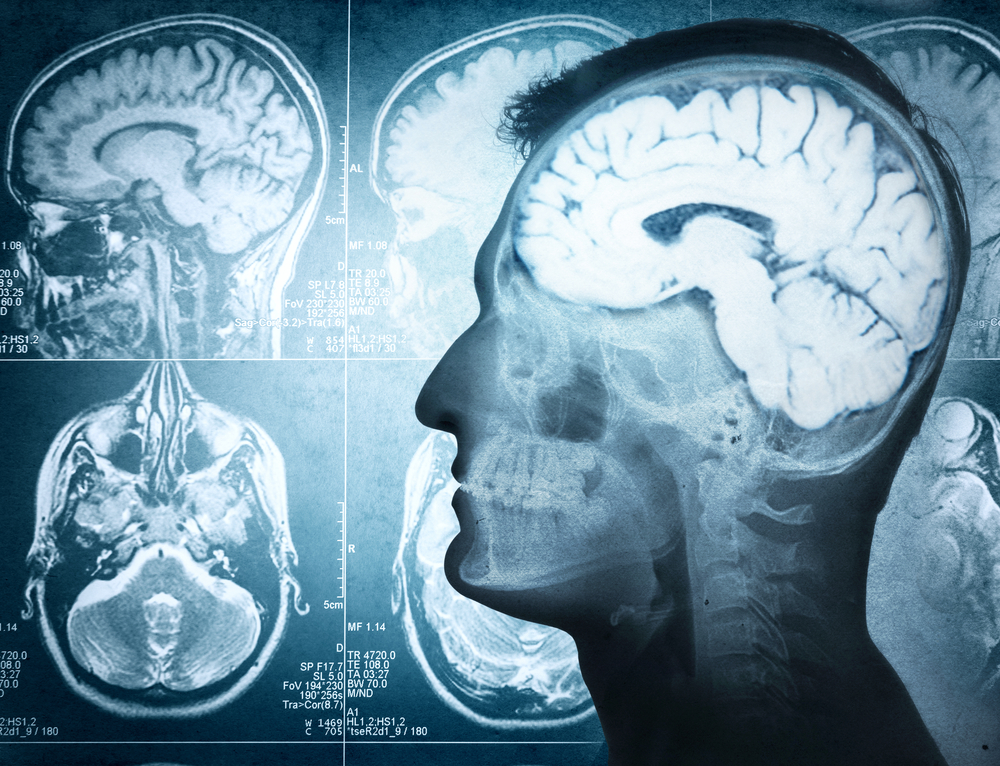
Researchers used magnetic resonance imaging (MRI) to analyze the brains of patients to gain insight into the mechanisms underlying their neurological problems.
The study titled “Cognitive and functional connectivity alterations in Friedreich’s ataxia” was published in the journal Annals of Clinical and Translational Neurology.
In addition to progressive neurological disability, people with Friedreich’s ataxia can develop a mild impairment of other neuropsychological functions including impaired emotion recognition, visual and spatial recognition, information processing speed, and memory.
A few studies looking at the brains of patients using MRI exams suggested that these symptoms may be due to the presence of deficits in the connectivity between different brain areas. But these studies were done while patients were performing a motor or a behavioral task.
In this study, scientists wanted to see how the brain of a person with Friedreich’s ataxia behaves when they are in a resting state to correlate the observations with neurological problems.
Using an imaging technique called functional magnetic resonance imaging (fMRI), researchers examined neuronal activity while the subjects were not performing any particular task.
The goal was to determine the connection between the activity of distinct brain regions during the individual’s resting state, a property known as functional connectivity.
The study included 24 Friedreich’s ataxia patients and 24 healthy controls with a mean age of 31.
All patients underwent a thorough clinical evaluation of neuropsychological symptoms, including a battery of tests assessing language, executive functions, memory, and visual perception.
Each individual’s brain was scanned by fMRI, focusing on brain areas potentially related to the patients’ compromised functions, namely the multiple subregions of the cortex. The assessment was done while patients were lying in a relaxed position.
Friedreich’s ataxia patients performed worse in several neuropsychological activities compared with healthy controls, including global cognitive function, spatial memory, visual perception, attention, and motivation tasks.
Clear-thinking ability, nonverbal intelligence, language, and working memory were not significantly different than healthy adults, however.
Brain fMRI images revealed that patients had significant alterations in the interaction between brain areas. In terms of neuron activity, some regions in the brain cortex interacted less, while others interacted more compared to healthy individuals.
The reduction in the connection between cortex subregions is in line with known alterations in the crosstalk between cortex and cerebellum — a region that controls movement, language, and attention — in these patients, researchers say.
Conversely, higher connectivity between other cortex areas possibly reflects a natural compensatory event occurring in the brains of patients to counteract neuronal deficits.
The researchers concluded that these results “in conjunction with clinical findings and neurofunctional tests, may shed new light … on the dynamics of brain plasticity in FRDA [Friedreich’s ataxia].”