CrCEST Imaging Tool May Detect Mitochondrial Function in Muscles
Written by |

Creatine chemical exchange saturation transfer or CrCEST, a metabolic imaging tool, may be a valuable technique for detecting mitochondrial function across muscles in people with Friedreich’s ataxia (FA), a new study found.
To date, CrCEST has not been used to assess mitochondrial capacity in people with FA.
“CrCEST MRI may be a useful biomarker of muscle-group-specific decline … that can be leveraged to track within-participant changes over time,” the researchers wrote, adding, “We envision continued development of this metabolic imaging tool.”
The study, “In vivo assessment of OXPHOS capacity using 3 T CrCEST MRI in Friedreich’s ataxia,” was published in the Journal of Neurology.
FA is a rare genetic condition caused by mutations in the gene that carries instructions for making frataxin. This protein is important for the proper functioning of mitochondria, organelles that produce the necessary energy for cellular processes. Its deficiency leads to a disruption in energy production within cells.
Mitochondrial function in FA is typically measured with 31P magnetic resonance spectroscopy, but this approach has limited resolution and cannot be used to assess muscle-specific mitochondrial function. Such assessments are essential, since distinct muscle types have different metabolic needs, and their mitochondrial activity also will be distinct.
CrCEST MRI is a technique that measures free creatine, a metabolite required for mitochondrial function, in muscles. By measuring free creatine with CrCEST, and how it changes with exercise, scientists can indirectly assess mitochondrial capacity in specific muscles.
This mitochondrial capacity is essentially a measure of the maximal capacity of a muscle to produce energy from oxygen in a given period of time. The higher that capacity, the healthier the muscle is. However, clinicians have not used CrCEST to assess mitochondrial capacity in FA patients.
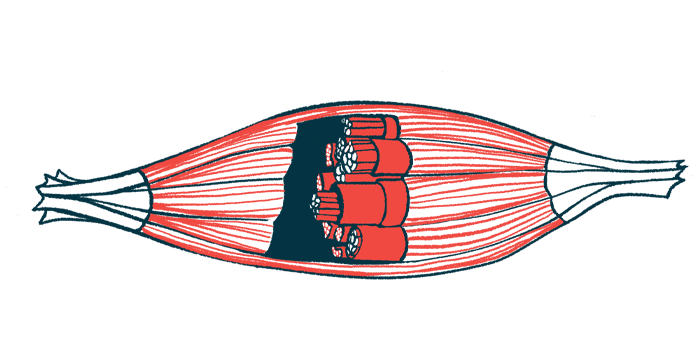
Now, scientists set out to compare mitochondrial capacity in the muscles of healthy adults and people with FA using CrCEST. They particularly focused on the calf muscle, which is made up of two main muscles — the gastrocnemius and the soleus. The gastrocnemius is further split into the medial gastrocnemius and the lateral gastrocnemius.
The team also evaluated the relationship between body composition, physical activity, and muscle mitochondria capacity.
All eligible participants completed questionnaires to assess their physical activity. The researchers measured each participant’s waist circumference, muscle mass, and fat mass. CrCEST MRI then was conducted on the participants’ calf muscles before and after plantar flexion exercise — a movement in which the top of the foot points away from the leg.
In all, 36 adults participated in the study: 11 with FA and 25 healthy people who served as controls. The mean age was 27 among the FA patients and 29 for the controls.
In total, 55% of patients and 40% of the controls had increased waist circumference, defined as one greater than 88 cm (34.6 inches) for females and 102 cm (48 inches) for males. The control group exercised for an average of 19.2 hours weekly, significantly more than the average of 11.1 hours of exercise among patients.
Pre-exercise free creatine levels were similar in all participants. But assessing each calf muscle group showed that creatine levels were higher in the soleus than in the lateral gastrocnemius, regardless of disease status.
After exercise, free creatine levels dropped. But while overall creatine levels changed similarly in both groups, people with FA used different muscles to complete the exercise, the data showed.
Specifically, the healthy controls used their lateral and medial gastrocnemius muscle more than the FA group, but all participants used the soleus muscle similarly.
“In the context of a plantar flexion exercise, individuals with FA with limited coordination and or strength may engage all muscle groups, while healthy individuals can efficiently perform the exercise using predominantly the most appropriately positioned muscle groups,” the researchers wrote.
“We observed that individuals with [FA] tended to engage all three muscles groups similarly, while healthy controls tended to use the gastrocnemius the most,” they added.
Free creatine levels tend to decrease after exercise. Overall, people who exercised more frequently had greater decreases in free creatine levels in the lateral gastrocnemius muscle after exercise. On the other hand, people with higher waist circumferences had smaller reductions in free creatine levels in the medial gastrocnemius and soleus muscles after exercise.
Compared with the control group, mitochondrial capacity after exercise was lower in the lateral gastrocnemius muscle in the FA group. However, when self-reported physical activity was taken into account, differences in mitochondrial capacity in the lateral gastrocnemius muscle were less evident.
“After accounting for differences in physical activity between [groups], differences in [mitochondrial capacity] were attenuated, highlighting challenges of detecting the differences between primary (i.e. genetic) and secondary (i.e., related to decreased mobility) adverse effects on muscle metabolism,” the researchers wrote.
Mitochondrial capacity also was lower in the medial gastrocnemius muscle in the FA group, although the difference was not statistically significant. Interestingly, there was a link between physical activity and mitochondrial capacity in the medial gastrocnemius for all participants.
“Notably, we found … [that] the effect of disease status on [mitochondrial function] … differs by muscle group,” the researchers wrote.
The researchers plan to carry out further studies and continue developing the CrCEST technology to accurately assess mitochondrial capacity in people with mitochondrial disorders.


