Patient Telomeres on DNA Involved in FRDA Pathogenesis
Written by |

Often times, researchers focus on cells from the brain or the heart when studying Friedreich’s ataxia. A new study from Brunel University London and University of Cambridge in the United Kingdom chose instead to focus on blood leukocytes and skin fibroblasts from patients with Friedreich’s ataxia. The reasoning was based on previous evidence that these cells have dysfunctional responses to oxidative stress.
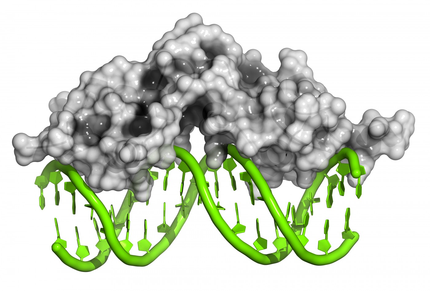
As reported in “Identification of Telomere Dysfunction in Friedreich Ataxia,” which was published in the journal Molecular Neurodegeneration, “We aimed to investigate both telomere structure and function in Friedreich ataxia cells.” Telomeres are the end caps to DNA that help maintain its integrity. Since telomeres are such a vital part of genomic stability, it is dangerous when they are damaged by oxidative stress that can occur when iron accumulates in cells (which happens in Friedreich’s ataxia due to frataxin protein deficiency).
To start investigating alterations in telomeres in patients with Friedreich’s ataxia, the researchers first measured the length of patient-derived fibroblasts’ and leukocytes’ telomeres. While fibroblasts had significantly longer telomeres in Friedreich’s ataxia patients than in healthy controls, leukocytes had significantly shorter telomeres. Going a step further and analyzing telomeres obtained from cerebellum autopsy tissues, the researchers identified that these cells also had shortened telomeres.
To explain the discrepancy between fibroblast telomere length and those of cerebellar tissue and leukocytes, the researchers looked at telomerase function. Their assay showed that telomerase was not even present in patient-derived fibroblasts, indicating that the protein that normally regulates telomere length was not involved. Instead, the researchers discovered that a process known as ALT was responsible and was significantly more involved when cells were in oxidative stress.
Rounding out the team’s experiments was a direct test for telomere dysfunction in patient-derived fibroblasts. “The presence of telomere dysfunction-induced focis (TIFs) in a cell is indicative of DNA damage at telomeres and is used to quantitatively measure levels of telomeric dysfunction,” explained the authors. “Friedreich ataxia cells had greater TIF frequencies in comparison to controls, indicating that Friedreich ataxia fibroblasts have greater induced telomere dysfunction.”
This study helps explain why cells are so affected by oxidative damage in patients with Friedreich’s ataxia. A lack of protection on the ends of DNA induces greater cell dysfunction, leading to tissue structures that cannot function as efficiently. By understanding telomere dysfunction and recognizing another biomarker of Friedreich’s ataxia, the researchers may help create another way of identifying the severity of disease in patients with Friedreich’s ataxia.


