Researchers spot new ultrasound patterns across peripheral nerves in FA
Ultrasound picked up changes in key arm and leg nerves in small study
Written by |


High-resolution nerve ultrasound revealed nerve enlargement, a sign of nerve changes, in the arms and legs of people with Friedreich’s ataxia (FA), a study in Germany shows.
The study also found that some people showed nerve atrophy, or shrinkage, in the arms and legs. Together, the findings highlight that peripheral nerve involvement can vary in FA. Peripheral nerves are the nerves outside the brain and spinal cord.
The study, “Nerve Ultrasound in Patients With Friedreich Ataxia,” was published in Muscle & Nerve.

What causes Friedreich’s ataxia and how it affects nerves
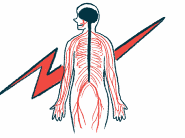
FA is caused by mutations in the FXN gene that reduce levels of frataxin, a protein needed for mitochondria, the cell’s energy-producing structures. Because muscle and nerve cells rely heavily on energy, low frataxin can contribute to FA symptoms such as coordination and balance problems (ataxia) and neurological issues.
Nerve enlargement, meaning a nerve is thicker than normal, has been reported in people with FA. While the cause is still unclear, researchers have suggested possible roles for inflammation and dysmyelination, changes in the myelin coating that helps nerves send signals.
In this study, researchers in Germany evaluated whether high-resolution ultrasound (HRUS) could help assess peripheral nerve changes in 20 people with FA. Participants included nine men and 11 women and had a mean age of 41.6 years. Symptoms began at a mean age of 17 years.
All participants had signs of sensory neuropathy, which can cause abnormal sensations or reduced feeling. Six had cardiomyopathy (heart muscle disease) and three had diabetes.
Of the 13 participants who had nerve conduction studies, 12 showed sensory neuropathy, but none had signs of demyelination on these electrical tests (meaning no evidence of slowed signal conduction typical of myelin loss).
Study detects mixed patterns across arm and leg nerves
All participants underwent HRUS, and changes were detected in 17 of the 20 patients (85%). Nerve enlargement was most often seen in the median and/or ulnar nerves in the upper arm (45%). Two of these patients also had nerve enlargement in the forearm and the cubital fossa (front of the elbow), though the difference was not statistically significant compared with normal nerve size.
In the legs, nerve enlargement was seen in the tibial, fibular, and sural nerves in 20% of patients, also without statistical significance. Overall, nerve enlargement was not linked to disease duration or severity.
Nerve atrophy, or shrinkage, was also found in the arms. Two patients showed atrophy in the median and ulnar nerves, and five had significant shrinkage of the ulnar nerve in the forearm.
In the legs, shrinkage of the tibial nerve was seen in six patients at the back of the knee and in another six at the inner ankle. Shrinkage of the fibular nerve at the back of the knee was found in two participants, but the difference was not statistically significant.
Both enlargement and shrinkage occurred in seven patients (35%), while five each (25%) had enlargement alone or shrinkage alone. In some cases, enlargement and atrophy were seen in different parts of the same nerve, “underscoring the [variable] nature of peripheral nerve involvement in [FA],” the researchers wrote.
The researchers wrote that it remains unclear whether these changes happen at the same time or follow a consistent pattern. “Our findings suggested an asynchronous [not at the same time] degeneration–regeneration cycle across different nerve segments,” they wrote. However, further studies are needed to “clarify the underlying [mechanisms] and guide potential treatment.”