Tests Reveal Cardiomyopathy Associated With Freidreich’s Ataxia


Patients with Friedreich’s ataxia are at a high risk for developing hypertrophic cardiomyopathy, where the walls of the heart become thicker than normal. Little is known about hypertrophic cardiomyopathy disease progression in Friedreich’s ataxia patients, although the condition can be severely life-limiting. To address this lack of knowledge, researchers at the Comprehensive Heart Failure Center at the University of Würzburg in Germany conducted a thorough evaluation of Friedreich’s ataxia patients with cardiomyopathy and discovered that a comprehensive cardiac assessment can detect the condition nearly 100% of the time.
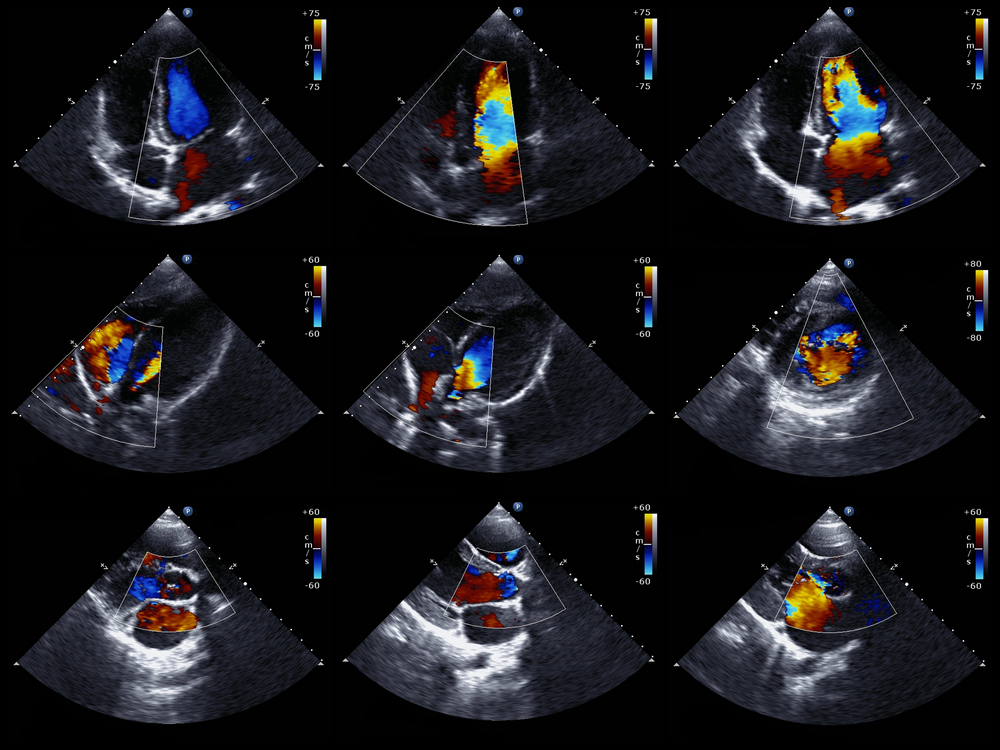
The new work entitled, “The Cardiomyopathy in Friedreich’s Ataxia — New Biomarker for Staging Cardiac Involvement,” was published in the International Journal of Cardiology. It builds off of the group’s work in 2012 that established a staging algorithm for describing cardiac involvement in Friedreich cardiomyopathy. In the present study, the researchers evaluated 32 patients with Friedreich’s ataxia using a variety of techniques including resting electrocardiogram, 24-hour Holter-electrocardiogram, echocardiography, and magnetic resonance imaging.
Each evaluation technique was conducted for a specific reason. Resting electrocardiogram was used to determine abnormalities in electrical signals in the heart, 24-hour Holter electrocardiogram was used to detect abnormal heart rhythms, and echocardiography and magnetic resonance imaging were used to visualize the heart. In addition to these evaluation techniques, the researchers also collected samples of blood to detect biomarkers in patients’ serum.
All but two of the 32 Friedreich’s ataxia patients had cardiomyopathy. Using the staging system of previous, the researchers stratified the patients into end-stage (25% of patients), severe stage (41% of patients), intermediate stage (13% of patients), and early stage (16% of patients) cardiomyopathies.
Using these stratifications, the researchers were able to correlate disease severity to heart wall thickness. They noticed that early-stage cardiomyopathies showed increased wall thickness, while more advanced disease showed wall remodeling and decreased thickness. “The current study suggests that a Friedreich patient should be evaluated at least once by sophisticated imaging and electrocardiography including blood biomarkers,” commented the authors. Although a comprehensive cardiac assessment can reveal cardiomyopathies in nearly 100% of Friedreich’s ataxia patients, patients with advanced fibrosis in the heart may be missed due to remodeling processes that affect wall thickness. Therefore, in order to “see” cardiomyopathies, it is important to both visualize the heart and test serum for biomarkers of disease.